| Authors |
Berry C, L'Allier PL, Gregoire J, Lesperance J, Levesque S, Ibrahim R, Tardif JC. |
|
| Title |
Comparison of intravascular ultrasound and quantitative coronary angiography for the assessment of coronary artery disease progression |
|
| Full source | Circulation 2007;115:1851-7 | |
|
|
Per scorrere le diapositive |
|
|
||
| Abstract |
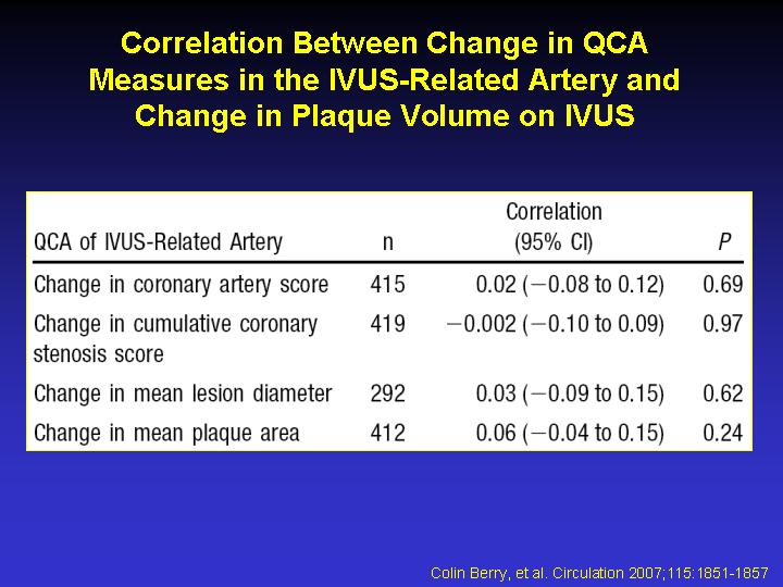
BACKGROUND: The relative merits of quantitative coronary analysis (QCA) and intravascular ultrasound (IVUS) for the assessment of progression/regression in coronary artery disease are uncertain. To explore this subject further, we analyzed the angiographic and IVUS data derived from a contemporary clinical trial population. METHODS AND RESULTS: We investigated the relationships between QCA and IVUS at single time points (n=525) and also for the changes over time (n=432). QCA and IVUS data underwent central laboratory analyses. Statistically significant correlations were observed between the QCA coronary artery score and the IVUS-derived lumen volume (r=0.65, P<0.0001) and total vessel volume (r=0.55, P<0.0001) and between the QCA cumulative coronary stenosis score and percent atheroma volume on IVUS (r=0.32, P<0.0001) at baseline for matched segments. A similar pattern of correlations was observed for global (all segments) QCA-derived and single-vessel IVUS-derived data. There were statistically significant but weak correlations between the changes over time in lumen dimensions on QCA and IVUS (P=0.005) and between the change in cumulative coronary stenosis score on QCA and percent atheroma volume on IVUS (r=0.14, P=0.01). Nevertheless, patients with and without angiographic progression had changes in plaque volume on IVUS of 9.13 and 0.20 mm3, respectively (P=0.028). CONCLUSIONS: QCA- and IVUS-derived measures of lumen dimensions are correlated at single time points and for changes over time. Although the change in percent atheroma volume is only weakly correlated with QCA changes as continuous variables, disease progression on QCA is associated with significant increases in plaque volume on IVUS compared with no angiographic progression.
|
|